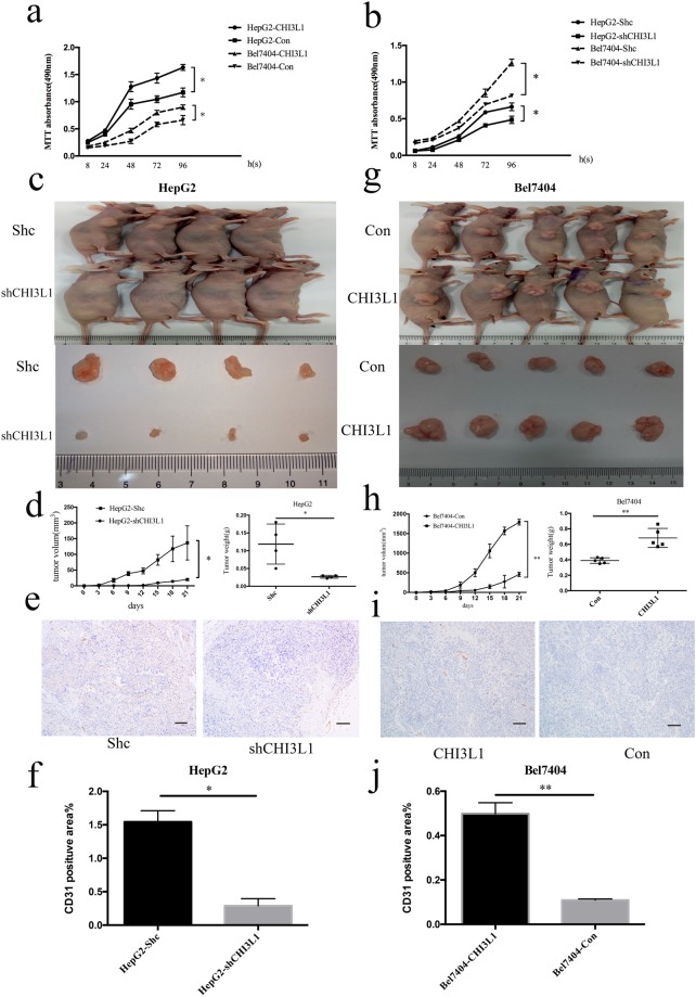
Figure 4.
CHI3L1 promotes cancer cell proliferation in vivo and in vitro. (a,b) CHI3L1 overexpression and knockdown cells and the control cells were seeded into 96 well plate. Cells were treated with MTT and the MTT absorbance value was measured at 8 h, 24 h, 48 h, 72 h and 96 h after cell seeding. (c,d,g,h) Representative images, growth and weight of tumors following subcutaneous injection of the HepG2-shCHI3L1, Bel7404-CHI3L1 or the control cells. (e,i,f,j) CD31 stain of the tumors formed by injection of the HepG2-shCHI3L1 and Bel7404-CHI3L1or the control cells. Scale bar represent 200 μm. * and ** represent p < 0.05 and p < 0.01 by two-tailed t test.

