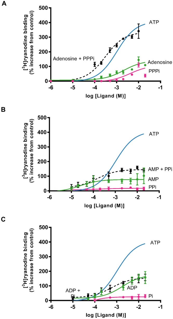
Figure 4.

Investigating the ability of ATP fragments to stimulate [3H]ryanodine binding as effectively as ATP alone. For clarity, in each graph, the ATP data from Fig. 3B is shown as a solid blue line. (A) Comparison of the effects of PPPi alone (pink), adenosine alone (green) and adenosine in the presence of 10 mM PPPi (black dashed line). (B) Comparison of the effects of PPi alone (pink), AMP alone (green) and AMP in the presence of 10 mM PPi (black dashed line). (C) Comparison of the effects of Pi alone (pink), ADP alone (green) and ADP in the presence of 10 mM Pi (black dashed line). Where combinations of ligands were used, they were added simultaneously at the start of the incubation period. The addition of Pi to ADP and PPi to AMP did not significantly increase binding above that observed with ADP (p = 0.2101) or AMP (p = 0.1917) alone whereas the addition of PPPi significantly increased binding above that observed with adenosine alone (**p < 0.01 (0.0065)). The error bars indicate SEM (n = 5). Where not shown, error bars are within the symbol.
