Fig. 4.
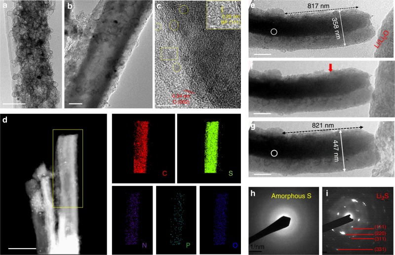
Integrate black phosphorus quantum dots with porous carbon/sulfur cathodes. a Transmission electron microscopy (TEM) image of the porous carbon nanofiber (PCNF) consisting of numerous hollow carbon spheres, b TEM image of the porous carbon host in a filled with sulfur particles and black phosphorus quantum dot (BPQD), designated as PCNF/S/BPQD, c high-resolution TEM (HRTEM) image showing the graphitic carbon layers of the PCNF and the uniform dispersion of BPQD within PCNF/S/BPQD, d scanning tunneling electron microscopy (STEM) image and energy dispersive spectroscopy (EDS) elemental mapping of the PCNF/S/BPQD fibers, e–g TEM images captured during lithiation of a PCNF/S/BPQD fiber, the red arrow in f refers to the reaction front, h, i selected area electron diffraction (SAED) patterns of the selected area with white circles in e and g, respectively. Scale bars, 100 nm (a); 200 nm (b); 10 nm (c); 2 nm inset (c); 500 nm (d); 200 nm (e–g)

