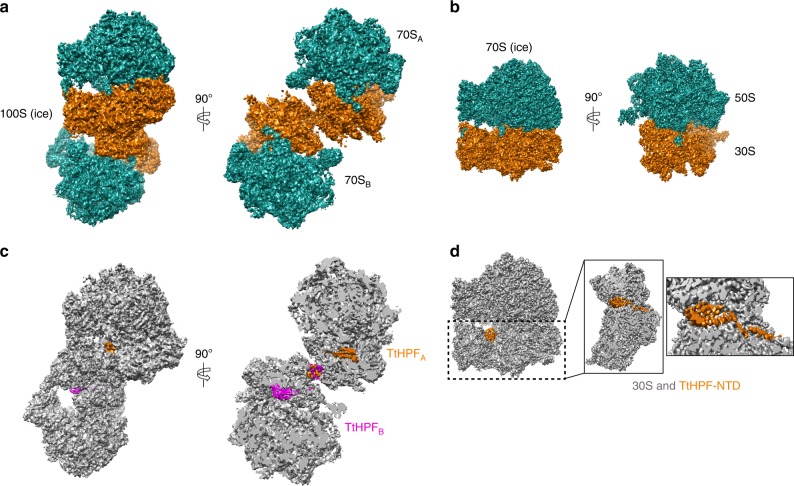
Fig. 2.
Cryo-EM structures of 100S (ice) and 70S (ice). a Orthogonal views of 100S (ice) with 50S subunits in green and 30S subunits in orange showing the two 70S copies constituting the 100S particle. b Orthogonal views of 70S (ice), coloring of subunits as in (a). c Views of 100S (ice) and slice-through view with both 70S ribosome copies colored in gray and the two TtHPF protein molecules colored in orange and magenta showing location of TtHPF-NTD and CTD within the 100S ribosome dimer. d View of 70S (ice) with TtHPF-NTD colored in orange. Close-up views on 30S subunit show location of TtHPF-NTD and the linker region. There was no density for the TtHPF-CTD in 70S (ice) reconstruction

