Figure 2.
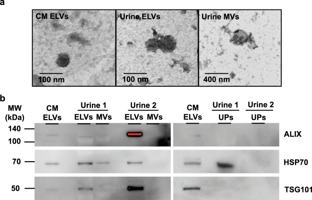
Validation of ELVs presence by particle and protein analyses. (a) Transmission electron microscopy (TEM) with the negative staining showed cup-shaped vesicles with a diameter less than 100 nm resembling exosomes presented in the ELV sample, whereas the vesicles larger than 100 nm were observed in the MV sample. Magnification power of 100,000x and 400,000x were applied for the MV and ELV samples, which corresponding to scale bars of 400 nm and 100 nm, respectively. (b) Western blot analysis showed enrichment of three exosome markers (i.e., ALIX, TSG101 and HSP70) in urinary ELVs as compared to urinary MVs and UPs isolated from two independent experiments. ELVs separated from culture media (CM) of MOLM13 cells served as the positive control in validation experiments. The red color in the immunoreactive band indicated the saturated signal intensity. The full-length blots of three exosome markers were provided in Supplementary Figure S1.

