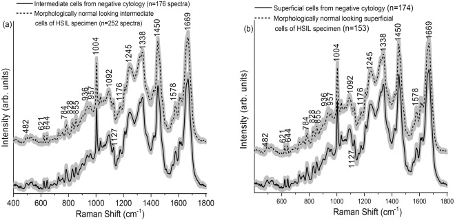
Figure 2.
(a) Mean Raman spectra ±1 standard deviation (SD) acquired from the intermediate cells of negative cytology ThinPrep specimens (n = 18) and morphologically normal appearing intermediate cells of high-grade squamous intraepithelial lesion (HSIL) ThinPrep specimens (n = 17). (b) Mean Raman spectra ±1 SD acquired from the superficial cells of negative cytology specimens (n = 18) and morphologically normal appearing superficial cells of HSIL cytology ThinPrep specimens (n = 17).

