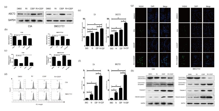
Fig. 2.
Resveratrol enhances cisplatin-induced toxicity via a DNA damage causing apoptosis-independent mechanism. (A) Western blot was performed to determine the expression levels of glutamine transporter ASCT2 in C3A and SMCC7721 cells after treated with indicated drugs. β-Actin was used as a loading control. (B) Resveratrol inhibits the uptake of glutamine in C3A and SMCC7721 cells. (C) Resveratrol decreases glutathione content in C3A and SMCC7721 cells. (D) Levels of ROS were measured by flow cytometric analyses in C3A and SMCC7721 cells. (E) Relative ROS levels in C3A and SMCC7721 cells are shown compared to control cells. (F) Quantification of γ H2AX immunofluorescent staining by IOD/Area of C3A and SMCC7721 cells. (G) Picture of γ H2AX immunofluorescent staining of C3A and SMCC7721 cells. (H) Western blott assays were performed to determine the expression levels of mitochondria and kytoplasm cytochrome c, caspase-9 and cleaved caspase-3 in C3A and SMCC7721 cells. β-Actin was used as a loading control. *P < 0.05, **P < 0.01, ***P < 0.0001.

