Figure 5.
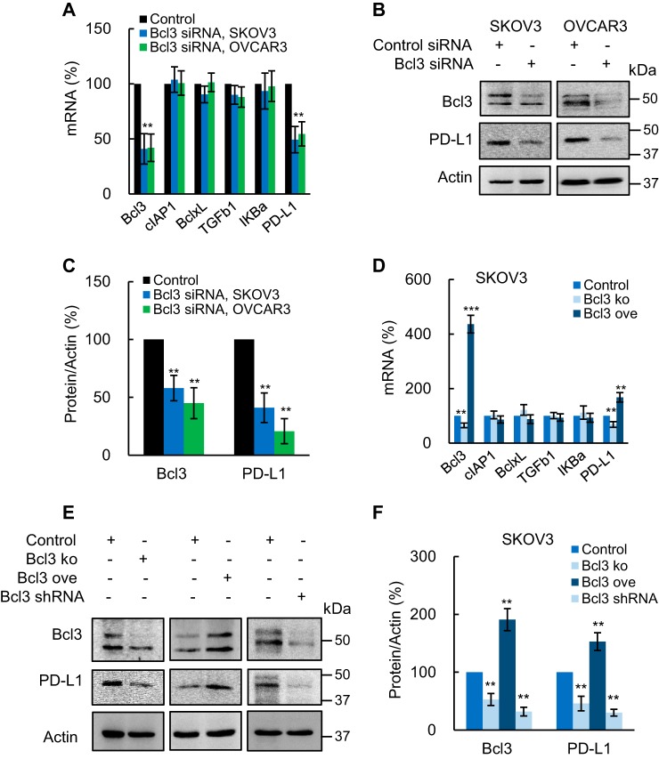
Bcl3 promotes constitutive PD-L1 expression in OC cells. A, expression of NF-κB-dependent genes measured by RT-PCR in SKOV3 and OVCAR3 cells transfected with control and Bcl3 siRNA. B, Western blotting of Bcl3, PD-L1, and control actin in WCE from SKOV3 and OVCAR3 cells transfected with control and Bcl3 siRNA. C, densitometric evaluation of Bcl3 and PD-L1 protein levels shown in B; Bcl3 and PD-L1 densities were normalized to actin, and expressed as % compared with cells transfected with the control plasmids. D, RT-PCR of NF-κB-dependent genes in SKOV3 cells transfected with control, Bcl3 KO, or Bcl3 ove plasmids. E, Western blotting of Bcl3 and PD-L1 in WCE from SKOV3 cells transfected with control, Bcl3 KO, Bcl3 ove plasmids, or stably transfected with Bcl3 shRNA. For cells stably transfected with Bcl3 shRNA, the same gel as in Fig. 4B was used and the membrane was re-probed with PD-L1 antibody (the same images for Bcl3 and actin were used as in Fig. 4B). F, densitometric evaluation of Bcl3 and PD-L1 protein levels shown in E; Bcl3 and PD-L1 densities were normalized to actin, and expressed as % compared with cells transfected with control plasmids. The values represent the mean ± S.E. of three experiments.

