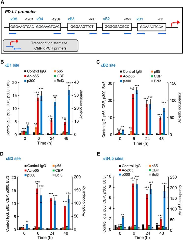
Figure 7.
IFN induces PD-L1 promoter occupancy by p65, Lys-314/315 acetylated p65, and p300 in OC cells. A, schematic illustration of NF-κB–binding sites in human PD-L1 promoter, and ChIP primers used in the ChIP assay. B–E, recruitment of p65, Lys-314/315 ac-p65, CBP, p300, and Bcl3 to PD-L1 κB1 (B), κB2 (C), κB3 (D), and κB4/κB5 sites (E) in IFN (50 ng/ml)-treated SKOV3 cells was analyzed by ChIP and quantified by real-time PCR; ChIP using control IgG is also shown. Each condition (antibody used at each time point) represents ∼1.25 × 10⁵ cells. The data are presented as fold-difference in occupancy of the particular protein at the particular locus compared with the human IGX1A (SA Biosciences) locus, and represent the mean ± S.E. of three experiments. Asterisks denote a statistically significant (*, p < 0.05; **, p < 0.01; ***, p < 0.001) change compared with ChIP using control IgG at the corresponding time.

