Figure 8.
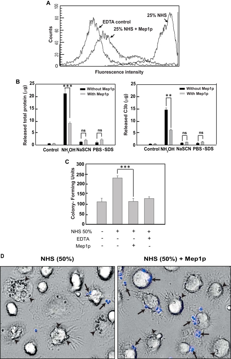
Mep1p inhibits C3b deposition on conidia and their phagocytosis. A, inhibition of C3b deposition by Mep1p. NHS (50%) was incubated with or without Mep1p (3 μg) in GVB2+ buffer for 60 min at 37 °C, and then this reaction mixture was further incubated with heat-inactivated conidia (1 × 10⁶). Control sample contained 20 mm EDTA. Bound C3b was detected by FACS using FITC-conjugated F(ab′)2 anti-C3 goat IgG. Mep1p considerably inhibited C3b deposition on conidia. B, C3b are bound to the conidial surface by ester linkage. Opsonized conidia were treated with NH2OH, NaSCN, or PBS/SDS, and the eluted proteins were estimated by Bradford assay (left panel) and ELISA (right panel; for details see the “Experimental procedures”) (**, p < 0.01; ***, p < 0.001). ns, not significant. In the case of opsonized conidia in the presence of Mep1p, the treatment of NH2OH released a significantly less amount of C3b. Mep1p inhibited covalent attachment of C3b on the surface of conidia. C and D, inhibition of phagocytosis by Mep1p. Conidia (1 × 10⁶) were opsonized by incubating with NHS (50%; 200 μl) for 60 min at 37 °C in the presence or absence of Mep1p (1 μg/200 μl 50% NHS), and the treated conidia were incubated with macrophages. Opsonizing conidia with NHS in the presence of Mep1p significantly decreased the conidial uptake by human monocyte-derived macrophages by at least 60% (***, p < 0.001). D, arrowheads indicate phagocytosed and arrows indicate adherent/nonphagocytosed conidia. The adherent/nonphagocytosed conidia were detected by calcofluor white staining. Mep1p treatment reduced the number of phagocytosed conidia.

