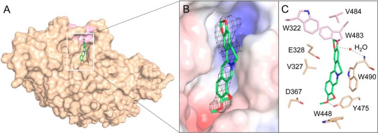
Figure 5.
Crystal structures of OfHex1 in complex with berberine. A, surface representations of OfHex1 complexed with berberine. B, binding mode of berberine in the active pocket of OfHex1. Electrostatic potential between −6 kT/e and 6 kT/e was shown as a colored gradient from red (acidic) to blue (basic). The 2Fo − Fc electron-density map around the ligand is contoured at the 1.0 σ level. C, amino acid residues involved in the binding of berberine in the active pocket of OfHex1. The hydrophobic recess accommodating the 1,3-dioxole group of berberine is shown in pink. The hydrogen bonds are shown as dashed black lines.

