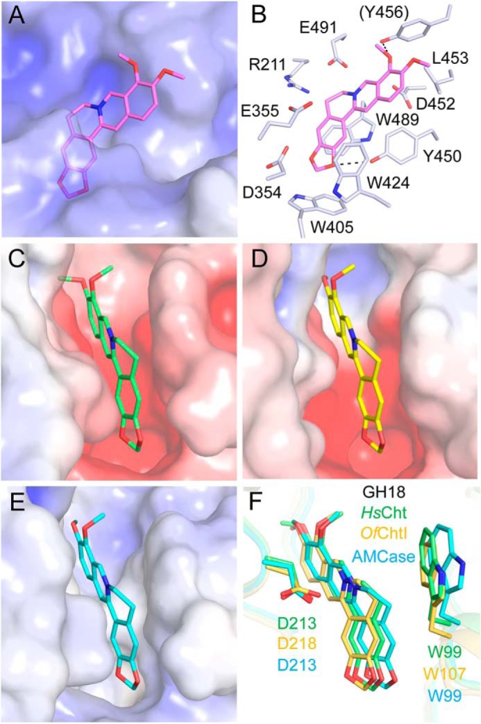
Figure 7.

Modeled structures of berberine in complex with GH20 and GH18 enzymes. Electrostatic potential between −6 kT/e and 6 kT/e was shown as a colored gradient from red (acidic) to blue (basic). A, binding mode of berberine in the active pocket of HsHexB. B, locations of the key residues for berberine binding in HsHexB. C, binding modes of berberine in the active pocket of HsCht. D, binding modes of berberine in the active pocket of OfChtI. E, binding modes of berberine in the active pocket of AMCase. F, superimposition of the binding modes of berberine with three chitinases. Residues of the HsCht and its berberine are shown in green. Residues of the OfChtI and its berberine are shown in yellow-orange. Residues of the AMCase and its berberine are shown in cyan.
