
Figure 5.
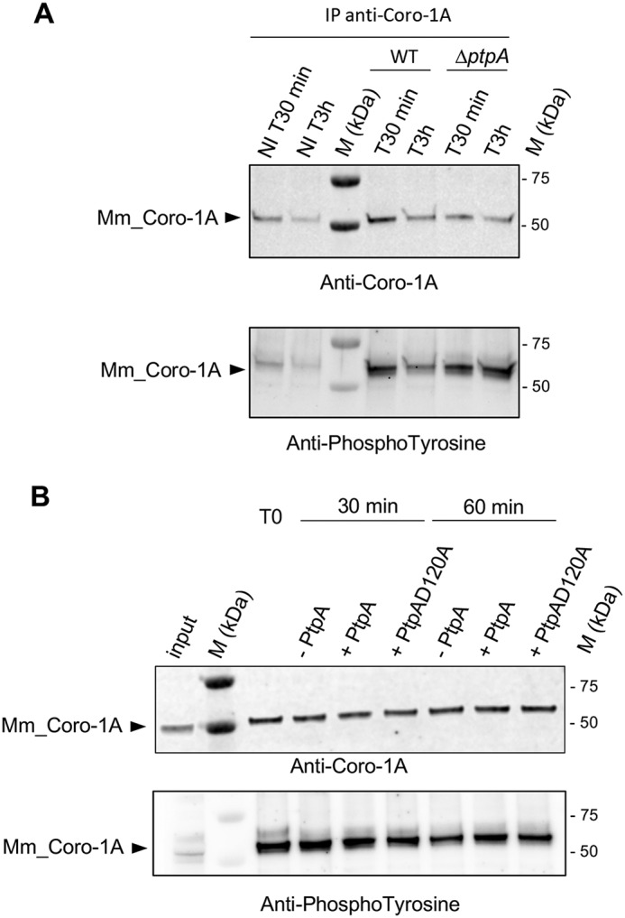
Coro-1A is phosphorylated on tyrosines in vivo but is not dephosphorylated by PtpA in vitro. A, immunoblot analysis with anti-Coro-1A (upper panel) or anti-phosphotyrosine antibodies (bottom panel) of immunoprecipitated (IP) endogenous Coro-1A (Mm_Coro-1A) from lysates of RAW 264.7 macrophages infected with cells of S. aureus Newman (WT) or the isogenic ΔptpA mutant. Noninfected macrophages and infected macrophages were treated with gentamicin for 30 min and subsequently incubated with lysostaphin to kill extracellular bacteria that might be released from lysed macrophages during the successive incubation time. Noninfected and infected macrophages were lysed 30 min (NI T30 min and T30 min) and 3 h (NI T3 h and T3 h) pGt treatment. B, immunoprecipitated Mm_Coro-1A from lysates of macrophages infected with strain Newman for 30 min pGt were incubated in the absence (−) or presence (+) of 2 μg of PtpA_His or PtpA-D120A_His at 37 °C for the time points indicated (T0, 30 and 60 min). Proteins were resolved by SDS-PAGE and probed with anti-Coro-1A (upper panel) or anti-phosphotyrosine (bottom panel) antibodies on the same blot. Contents of Mm_Coro-1A and Tyr-phosphorylated Mm_Coro-1A in lysates of S. aureus-infected macrophages prior to concentration by IP are indicated in the input lane (input). M kDa, molecular markers.