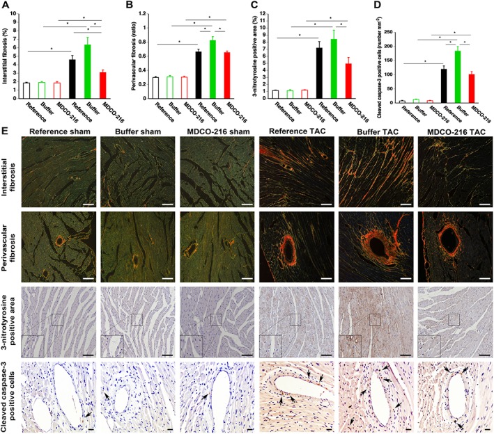
Figure 6.

MDCO‐216 infusion significantly reduces interstitial fibrosis, perivascular fibrosis, oxidative stress and cleaved caspase‐3‐positive cells in the myocardium after TAC. Sham mice and TAC mice are indicated by open and closed bars respectively. Bar graphs illustrating the degree of interstitial fibrosis (A), the degree of perivascular fibrosis (B), the percentage of 3‐nitrotyrosine‐positive area in the myocardium (C) and the cleaved caspase‐3‐positive cells in the myocardium (D) in reference sham mice (n = 11), buffer sham mice (n = 12), MDCO‐216 sham mice (n = 10), reference TAC mice (n = 21), buffer TAC mice (n = 17) and MDCO‐216 TAC mice (n = 21) at day 56 (reference mice) and at day 65 (buffer mice and MDCO‐216 mice) after sham or TAC operation. All data represent means ± SEM. *P < 0.05. Panel (E) shows representative photomicrographs of Sirius red‐stained interstitial collagen viewed under polarized light, of perivascular collagen viewed under polarized light, of myocardium immunostained for 3‐nitrotyrosine and of myocardium stained for cleaved caspase‐3. Scale bar represents 50 μm. Insets show a 4× magnification of the boxed region. Arrows indicate cleaved caspase‐3‐positive cells.
