
Figure 1.
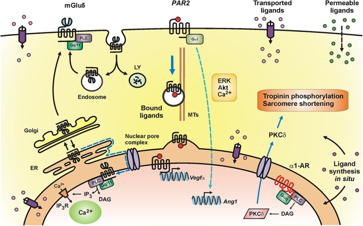
Schematic representation of selected intracellular GPCRs and ways in which they are activated. Left: proposed model of mGlu5 receptor trafficking in neurons in which >90% of mGlu5 receptors traffick through the golgi (Sergin et al., 2017). From there, between 15 and 40% (Hubert et al., 2001; López‐Bendito et al., 2002; O'Malley et al., 2003) go to the cell surface where they undergoe a cycle of constitutive endocytosis and recycling (Trivedi and Bhattacharyya, 2012). Alternatively, 60–85% of mGlu5 receptors are retrogradely trafficked back to the ER and via lateral diffusion (dotted blue line) reach the NM (Vincent et al., 2016; Sergin et al., 2017). Middle: ligand‐bound PAR2 can translocate from the retinal ganglion cell surface to the nucleus via importin‐β, Snx11 and dynein; nuclear PAR2 activates Vegfa expression. In contrast, signalling from cell surface PAR2 results in angiopoietin 1 expression (Joyal et al., 2014). Right: the https://www.google.com/url?sa=t&rct=j&q=&esrc=s&source=web&cd=3&cad=rja&uact=8&ved=0ahUKEwiDsp6cqrvVAhWr64MKHWP9AHIQFgg7MAI&url=https%3A%2F%2Fwww.ncbi.nlm.nih.gov%2Fpmc%2Farticles%2FPMC3930413%2F&usg=AFQjCNEaQB5359zmSlQuF092N7FE4mEbxAceptor (α1‐AR) is localized to the inner NM of adult cardiac myocytes where it is activated by noradrenaline transported via the http://www.guidetopharmacology.org/GRAC/FamilyDisplayForward?familyId=196#1021 located on the cell surface and ER membranes. The nuclear α1‐adrenoceptor couples to Gαq and PLC, which activate nuclear PKCδ. PKCδ is then transported out of the nucleus through the nuclear pore complex. PKCδ induces phosphorylation of troponin 1 to regulate contractility and ERK to regulate survival signalling (Wu et al., 2014; Wu and O'Connell, 2015). Intracellular GPCRs can be activated via (i) channels, transporters or exchangers recognizing specific ligands such as glutamate in the case of mGlu5 receptors or OCT3 for noradrenaline activation of α1‐adrenoceptors; (ii) alternatively, ligands sufficiently permeable can freely diffuse across cell membranes; (iii) as in the case of PAR2, receptor‐bound ligands can simply be internalized with a given GPCR; and (iv) ligands can be synthesized within the cell and either diffuse or be trafficked to a given cellular compartment.