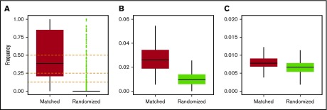
Figure 1.
Measurement of genetic similarity. Frequency distributions of IBD segments normalized by the total lengths of regions of interest for the following: (A) MHC, including HLA-A, HLA-B, HLA-C, HLA-DR, and HLA-DQ; (B) chromosome 6; (C) and the whole genome. Horizontal black bars represent median values. Outliers (green dots) are shown only for panel A (see text for details).

