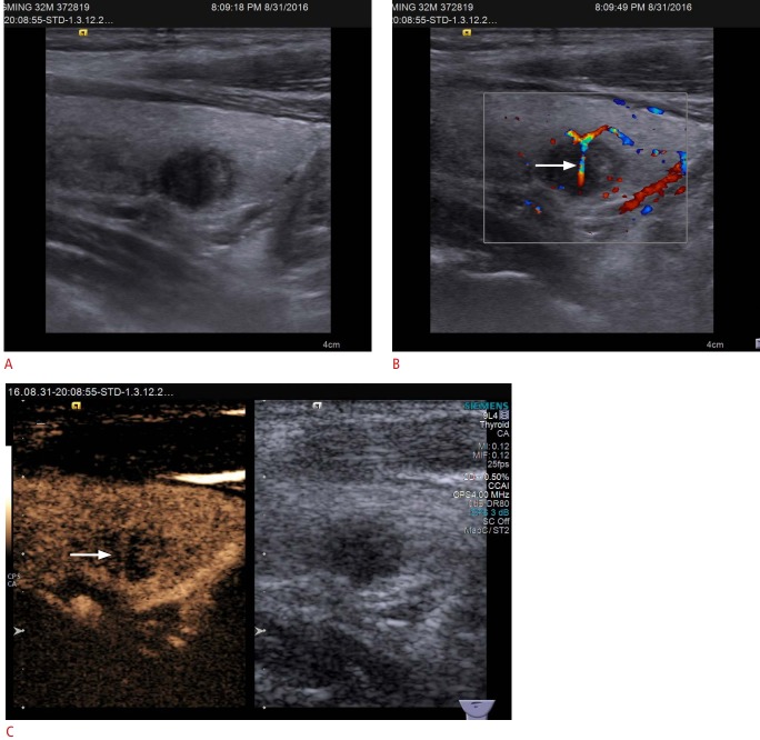
Fig. 1. Conventional ultrasonography (US) and contrast-enhanced ultrasound (CEUS) imaging of papillary thyroid carcinoma in a 32-year-old man.
A. Gray-scale US shows a hypoechoic solid nodule in the right thyroid lobe with a poorly defined margin, assessed as Thyroid Imaging Reporting and Data System category 4C. B. Color Doppler US shows branching vessels (arrow) within the mass. C. CEUS shows hypoenhancement with penetrating vessels (arrow) in the mass.

