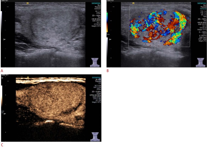
Fig. 3. Conventional ultrasonography (US) and contrast-enhanced US (CEUS) imaging of thyroid adenoma in a 35-year-old woman.
A. Gray-scale US shows an isoechoic nodule in the left thyroid lobe with a well-defined margin, assessed as Thyroid Imaging Reporting and Data System category 3. B. Color Doppler US shows mainly interior blood flow, with absent peripheral blood flow. C. CEUS shows homogeneity and ring enhancement in the mass.

