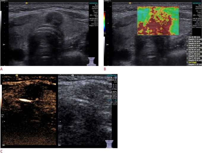
Fig. 5. Conventional ultrasonography (US), acoustic radiation force impulse imaging (ARFI), and contrastenhanced US (CEUS) imaging of thyroid adenoma in a 35-year-old man.
A. Gray-scale US shows a hypoechoic solid nodule in the isthmus with capsule contact assessed as Thyroid Imaging Reporting and Data System category 4C. B. ARFI is mainly red and slightly yellow, and the median shear wave velocity was 7.82 m/sec, which means that the nodule was very hard. C. CEUS shows hypoenhancement in the mass.

