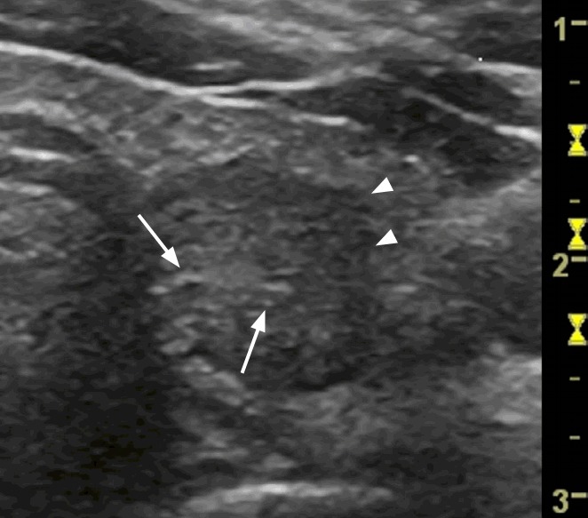
Fig. 2. A papillary carcinoma (confirmed by fine-needle aspiration cytology and surgery) in the left thyroid lobe of a 24-year-old woman.

A transverse ultrasonography shows a single, hypoechoic, solid nodule with irregular margins (arrowheads), containing some fine areas of hyperechogenicity (arrows) without any comet-tail artifacts that grew by >20% in the preceding 6 months. The total malignancy score of this nodule was 8.
