
Fig. 3. A follicular adenoma (the Bethesda System for Reporting Thyroid Cytopathology category III on fine-needle aspiration cytology) in the right thyroid lobe of a 76-year-old woman.
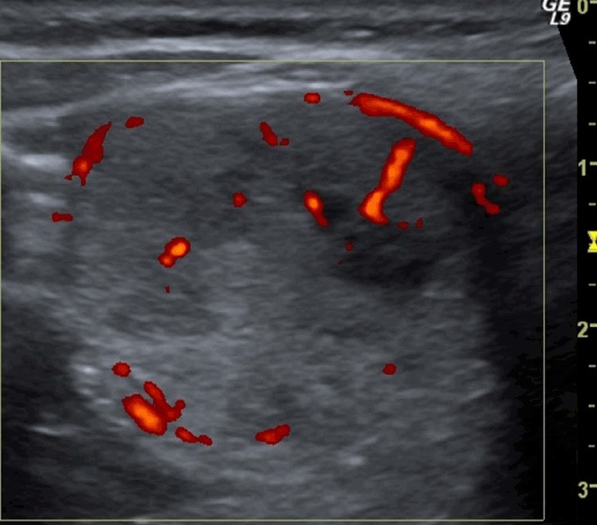
A transverse ultrasonography shows a single solid nodule (shown in the square) with mixed vascularization on a power Doppler examination that grew by >20% in the preceding 6 months. The total malignancy score of this nodule was 5.