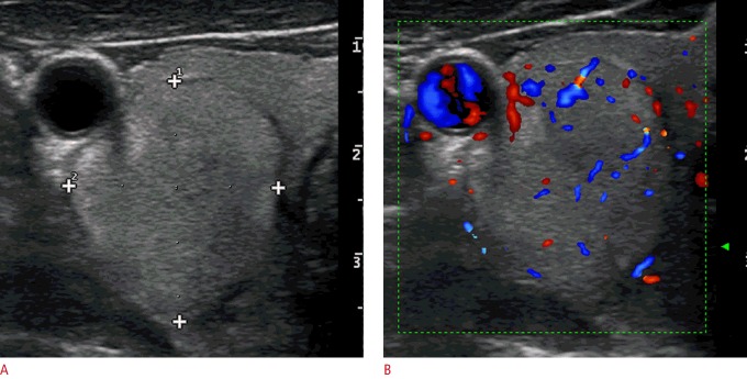
Fig. 5. A follicular adenoma (confirmed at surgery, follicular proliferation on fine-needle aspiration cytology) in the right thyroid lobe of a 47-year-old woman.
A, B. A transverse ultrasonography shows a single, isoechoic, thyroid nodule with regular margins, a taller-than-wide shape (A, anteroposterior diameter, 23 mm; latero-lateral diameter, 19 mm and B, a peri-intranodular Doppler signal). The total malignancy score of this nodule was 4.

