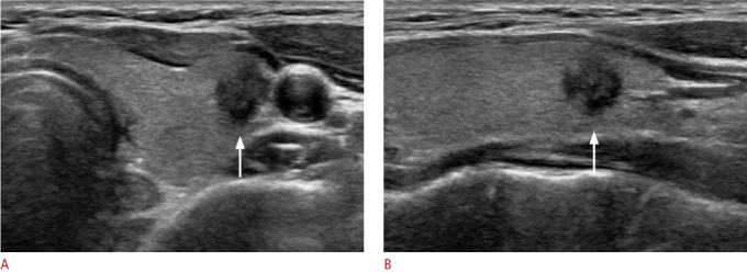
Fig. 2. A 46-year-old woman with conventional papillary thyroid carcinoma that was initially diagnosed as Bethesda category V (suspicious for malignancy) by fine-needle aspiration.
A, B. Ultrasonography (A, transverse; B, longitudinal view) shows a hypoechoic, irregular, non-parallel mass with spiculated margins in the left thyroid gland (arrows). The pathologic tumor size was 10 mm, and there was no evidence of lymph node metastasis or gross extrathyroidal extension at surgery.

