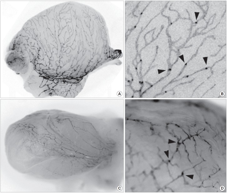
Fig. 1.
Visualization of the bladder lymphatic networks using fluorescent lymphatic reporter mouse and rat. Bladders were isolated from adult Prox1-EGFP mouse (A, B) and rat (C, D), and subjected to fluorescent stereomicroscopy. Fluorescent signals were captured using a monochrome camera and shown in grayscale. (A, C) Gross images revealed a dense network of lymphatic vessels on the surface of the bladders. (B, D) Enlarged images demonstrate lymphatic vessels and sprouts along with luminal valves (arrowheads).

