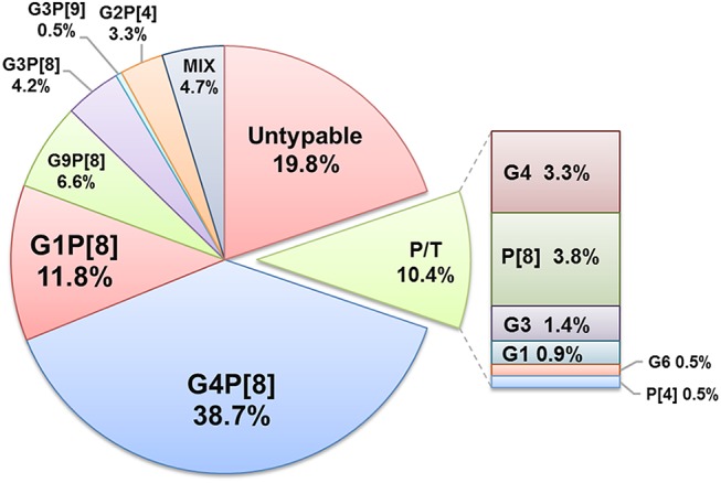
Fig. 3.

Distribution of RVA G/[P]-genotypes detected in clinical samples between 2009 and 2014 in Moscow. Each segment represents the relative distribution of an RVA G/[P] genotype based on data from type-specific multiplex real-time PCR analysis of fecal extracts collected from children with rotaviral enteritis (n = 212). The total figures for the whole period are presented. Mix mixed infection, P/T partially typed.
