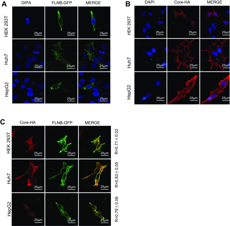
Fig. 2.

Filamin B colocalizes with core protein. (A) Confocal analysis of filamin B subcellular localization is shown in green. Cells were transfected with 2 μg FLNB-GFP plasmid. At 24 h post transfection, fixed cells were permeabilized and stained with DAPI. (B) Confocal analysis of core protein subcellular localization is shown in red. Cells were transfected with 2 μg of Core-HA plasmid. At 24 h post transfection, fixed cells were permeabilized and immunostained for core protein using anti-HA antibodies and fluorescent secondary antibodies. (C) Immunofluorescence analysis of the colocalization of filamin B and core protein is shown. Cells were transfected with 1 μg FLNB-GFP plasmid and 1 μg Core-HA plasmid. At 24 h posttransfection, fixed cells were permeabilized and immunostained for core protein using anti-HA antibodies and fluorescent secondary antibodies. Filamin B is shown in green (left), core protein in red (center), and colocalization in yellow (right). Scale bars represent 10 μm. Colocalization between filamin B and core protein was quantified using Leica confocal microscope software. R values are the Pearson correlation coefficient ± SD averaged from 5 to 8 cells.
