Figure 1.
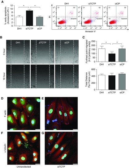
Silencing of translationally controlled tumor protein (TCTP) affected the apoptosis, migration, and morphology of blood outgrowth endothelial cells (BOECs). BOECs were transfected with DharmaFECT1 (DH1) alone, siTCTP, or nontargeting siRNA control (siCP) for 48 hours. (A) Apoptosis was assessed by flow cytometry using AnnexinV+ (FITC)/PI− (PE-Cy7-A) staining (n = 3; one-way ANOVA). (B) Cell migration was assessed using Ibidi culture inserts. After 24 hours, the inserts were removed and cells were treated with EBM-2 (2% FBS, 20 mM HEPES, and 2 μM hydroxyurea). Time-lapse microscopy was undertaken using a Leica SPE imaging system, with images taken every 5 minutes for 18 hours at 37°C. Representative images of BOECs at time 0 and 18 hours after wound healing are shown. (C) Quantification of the farthest point migrated and total distance migrated (n = 4; one-way ANOVA). (D and E) Representative confocal images of BOECs (D) untransfected or (E) transfected with siTCTP and stained with an antibody for TCTP (green) before counterstaining for F-actin (phalloidin; red) and nuclei (DAPI; blue). (F and G) Representative confocal images of BOECs (F) untransfected or (G) transfected with siTCTP and stained with an antibody for TCTP (green) before staining for α-tubulin (red) and counterstaining the nuclei (DAPI; blue). Scale bars: 25 μm. *P < 0.05. Error bars represent mean ± SEM.

