Figure 3.
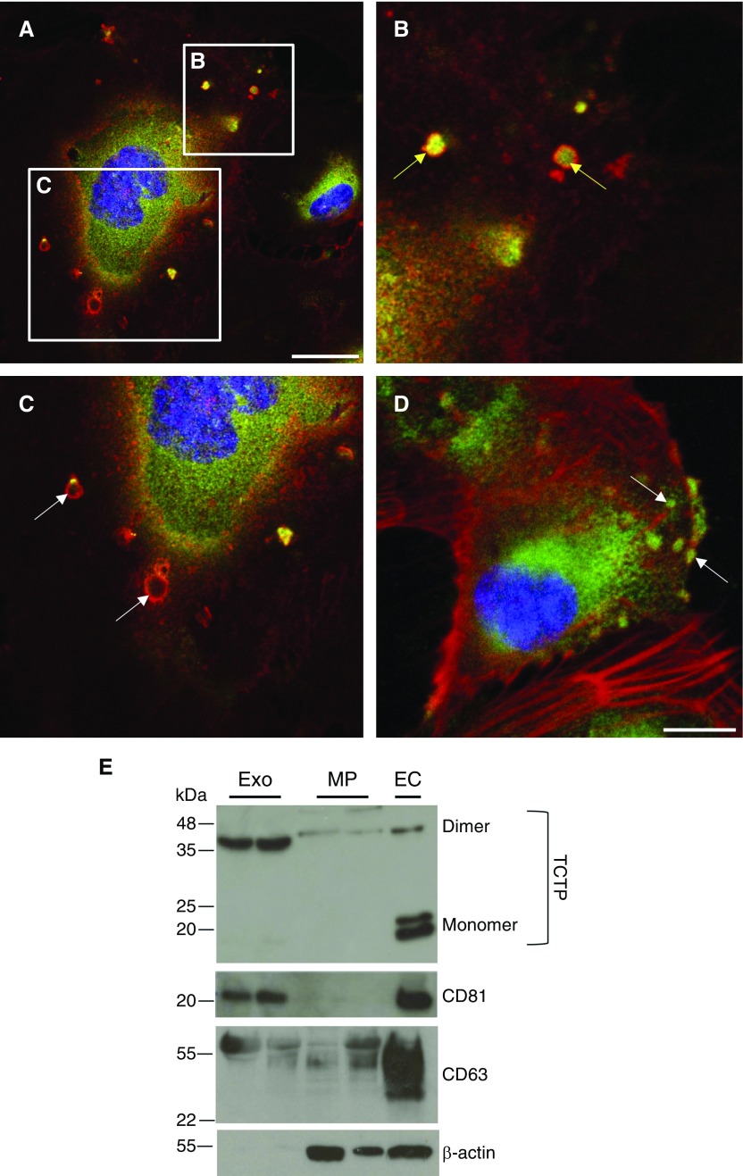
TCTP is exported via exosomes. (A–C) Representative confocal images of control BOECs stained using a TCTP antibody (green) and C81 (red). (A) BOECs secreting small particles (exosomes) positively stained for CD81 at the membrane. Scale bar: 25 μm. (B) Inset area in image A. Yellow arrows indicate exosomes positive for CD81 surface expression (red) and TCTP (green) intravesicle expression, with colocalization in yellow. (C) Inset area in image A. White arrows indicate TCTP-empty exosomes. (D) Representative confocal image of control BOECs stained using a TCTP antibody (green) and counterstained for F-actin (phalloidin; red). Scale bar: 25 μm. Localization of TCTP-positive intracellular vesicles (green) is indicated by white arrows. (E) Representative immunoblots for TCTP, CD81, CD63, and β-actin protein expression in exosomes (Exo), microparticles (MP), and BOECs (EC).

