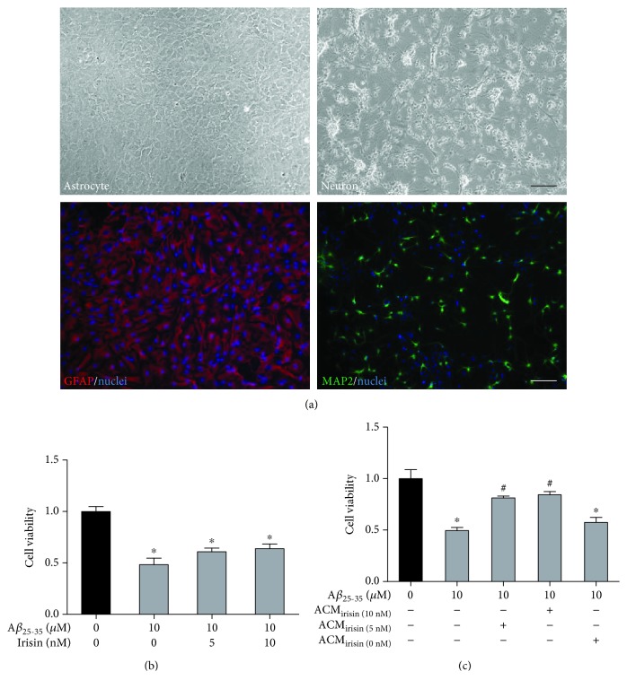
Figure 1.
Aβ25–35 caused significant loss of cell viability in primary cultures of hippocampal neurons, which is not reversed by irisin cotreatment. (a) Representative phase contrast photographs of cultured hippocampal astrocytes (2 weeks old) and neurons (7 days). Representative immunofluorescent photographs of astrocyte markers GFAP and MAP2. Hoechst 33258 used as nuclei staining. Scale bar represents 100 μM. (b) Irisin had no direct protective effects on the cell viability in neuron exposed to Aβ25–35 (MTT). (c) ACMirisin exerted protective effects on the cell viability in neuron exposed to Aβ25–35 while control ACM without pretreatment of irisin had no such protective effects (MTT). Data are expressed as means ± SEM, ∗p < 0.05 vs control, #p < 0.05 vs Aβ25–35, n = 6.

