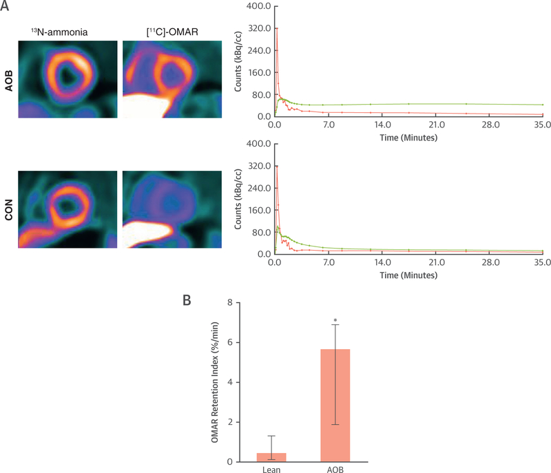
FIGURE 7. Myocardial Cannabinoid Type 1 Receptor Imaging Study in Humans.
(A) Short-axis positron emission tomographic/computed tomographic image (top left) of the left midventricular myocardium using the cannabinoid type 1 receptor (CB1-R) ligand [11C]-OMAR in a healthy subject with advanced obesity (AOB). Corresponding myocardial kinetics of [11C]-OMAR (top right) with time-activity curves (TACs) for arterial blood pool (pink) and myocardium (green). Furthermore, short-axis positron emission tomographic/computed tomographic image of the left midventricular myocardium in a healthy lean participant as a control subject (CON) (bottom left) and corresponding TAC (bottom right). Also here, there is relatively higher [11C]-OMAR myocardial uptake in a subject with AOB compared with lean CON that is mirrored by corresponding myocardial TAC course. (B) Quantitative analysis of myocardial OMAR retention index in humans with normal weight or CON and in subjects with AOB (n = 5 and n = 7, respectively); *p ≤ 0.05.

