Figure 2:

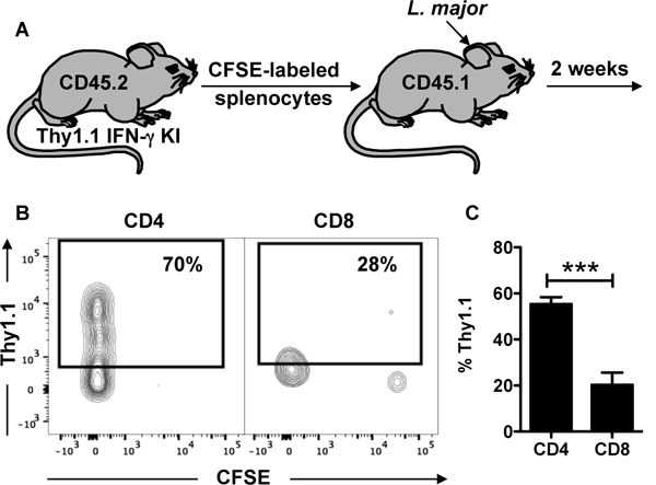
CD8+ T cells that have proliferated in response to L. major infection do not produce IFN-g in the skin. (A) Splenocytes from CD45.2 Thy1.1 IFN-g reporter mice were stained with CFSE and transferred into CD45.1 congenic mice infected with L. major. Two weeks post infection, mice were euthanized and donor cells were analyzed for CFSE dilution and IFN-g production. Depicted are (B) representative contour plots and (C) bar graph of Thy1.1 expressing donor CD4+ and CD8+ T cells. Flow plots pregated on live/singlets/CD3/CD8b or CD4. Representative data from 4 independent experiments (n = 3 mice) are presented. ***p ≤ 0.001
