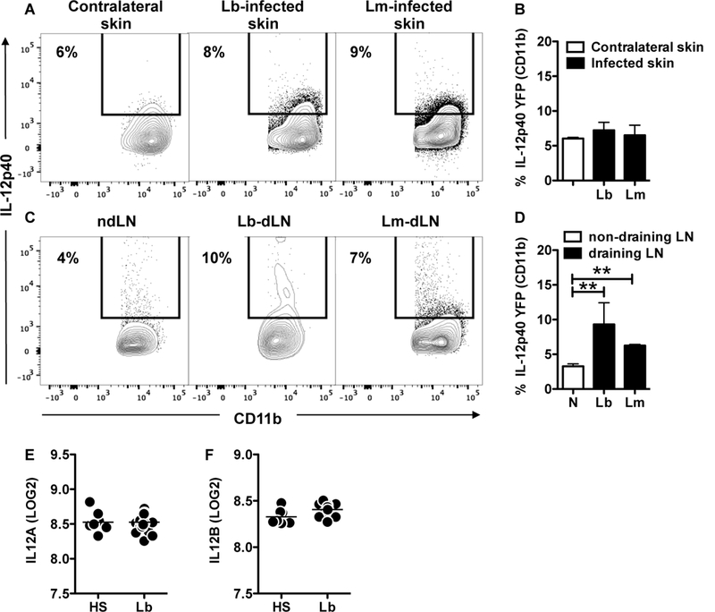
Figure 3:

IL-12 is not produced in leishmania lesions from mice and humans. IL-12p40 reporter mice were infected in the skin with L. major or L. braziliensis and 2 weeks post infection mice were euthanized. Cells from the (A, B) contralateral (a combination between contralateral skin from Lb and Lm infected mice) and infected skin or (C, D) non-draining (ndLN, a combination between ndLN from Lb and Lm infected mice) and draining lymph nodes (dLN) were analyzed for IL-12p40 expression directly ex vivo by flow cytometry. Depicted are (A, C) representative contour plots and (B, D) bar graphs showing the percentage of IL-12p40+ CD11b cells. Flow plots pregated on live/singlets/CD11b.Representative data from 2 independent experiments (n = 3 mice per group) with similar results are presented. **p ≤ 0.01. LOG2 expression of (E) IL12A and (F) IL12B in the skin of healthy subjects (HS) and L. braziliensis patients (Lb). Data obtained from 10 HS and 25 Lb.
