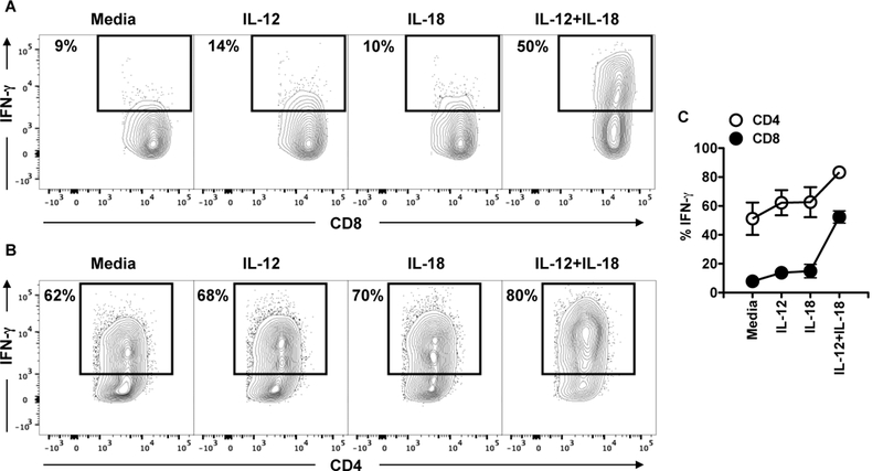
Figure 4:

CD4+ and CD8+ T cells have different requirements for IFN-g in leishmania-infected skin. C57BL/6 were infected in the skin with 106 L. major and 2 weeks post infection mice were euthanized. Cells from the infected skin were cultured with media or cytokines overnight and BFA for the last 4 hours; the expression of IFN-g was measured by flow cytometry. CD4+ and CD8+ T cells were analyzed for the expression IFN-g by flow cytometry. Depicted are (A and B) representative contour plots and (C) graph showing expression of IFN-g. Representative data from 4 independent experiments (n = 3 mice per group) with similar results are presented.
