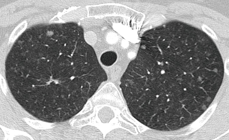
Figure 13.

Transverse thin CT section of the upper lobes shows multiple and bilateral subsolid nodules of variable size. A nodule of greater size and density is identified in the right upper lobe (arrow). In the context of multiple pulmonary nodules the recommendations is to assess the risk based on that of the largest nodule. Follow-up CT is recommended in 3–6 months.
