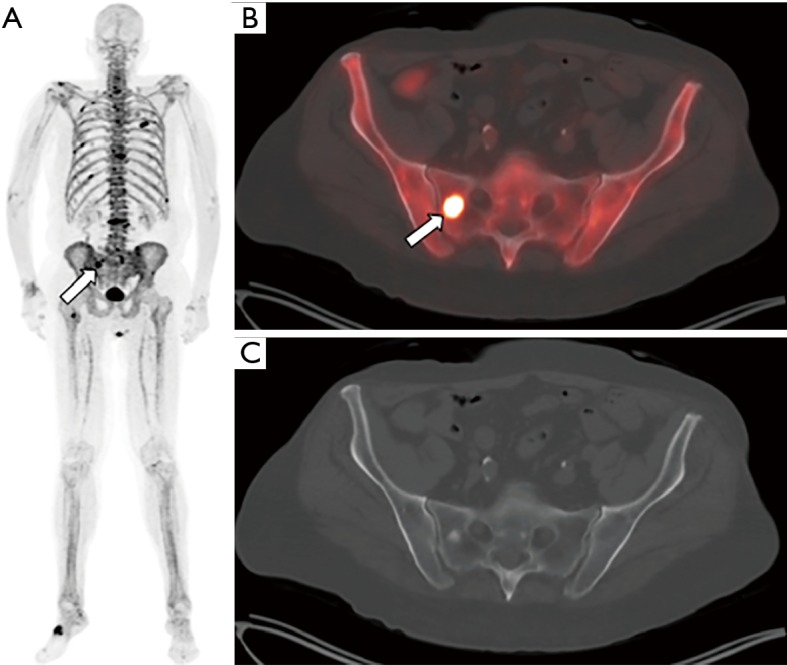
Figure 1.

Sodium [18F]fluoride-PET/CT images from a patient with prostate cancer status post external beam radiation therapy, androgen-deprivation therapy, and brachytherapy presenting with elevated PSA and biochemical recurrence. The maximum intensity projection image (MIP, panel A) demonstrates numerous foci of increased tracer activity in several ribs and the bony pelvis. The fused PET/CT (B) and CT only (C) images demonstrate a right sacral metastasis (arrow) with a sclerotic correlate.
