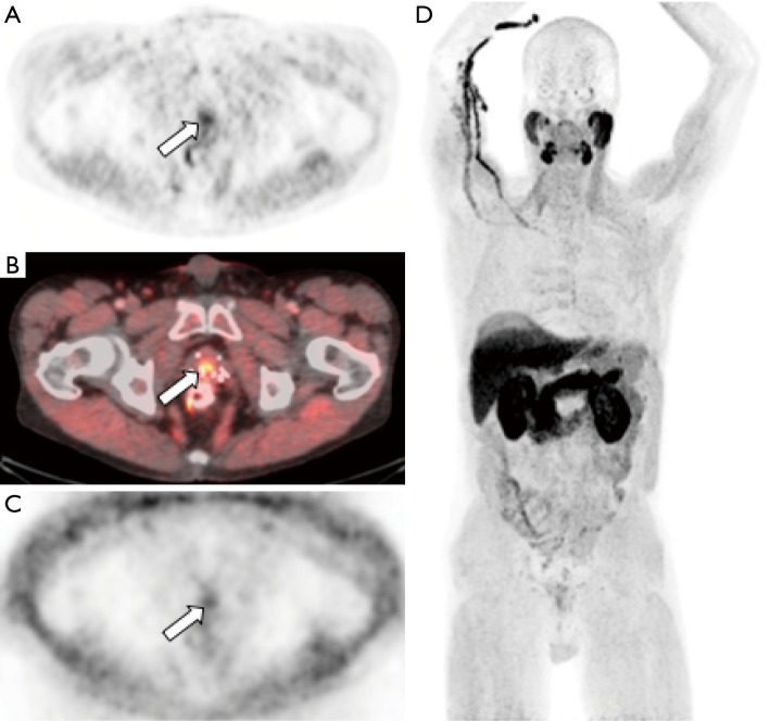
Figure 2.
[11C]choline-PET/CT images from a patient with prostate cancer status post brachytherapy, now with elevated PSA. Attenuation corrected PET (A) and fused PET/CT (B) images demonstrate focal tracer activity within the prostate gland (arrow) suspicious for recurrent tumor within the gland. Non-attenuation corrected PET images (C) demonstrate that the activity is not due to attenuation correction artifact from adjacent brachytherapy seeds. Whole body MIP images (D) demonstrates no additional distant metastases. Patient subsequently underwent prostatectomy which confirmed recurrent prostate cancer.

