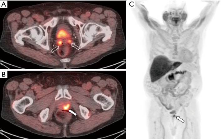
Figure 3.
Fused [18F]fluciclovine-PET/CT images (A and B) and whole body MIP image (C) from a patient with prostate cancer initially treated with radiation therapy and androgen-deprivation therapy reaching a PSA nadir of 0.7 ng/mL. The patient’s PSA continued to rise and measured 5.0 ng/mL at time of imaging which demonstrated local recurrence within the prostate gland (filled arrow) and invasion of the bilateral seminal vesicles (open arrows).

