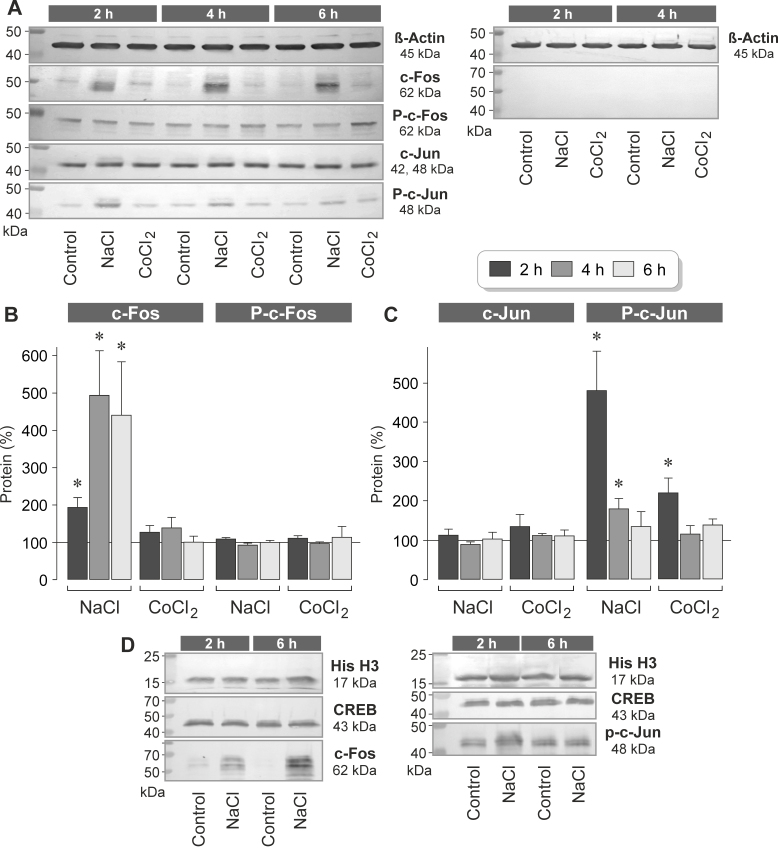
Figure 3.
High extracellular NaCl induces elevation in the levels of c-Fos and phosphorylated c-Jun proteins in RPE cells. The cells were stimulated for 2, 4, and 6 h with high (+ 100 mM) NaCl or the hypoxia mimetic CoCl2 (150 µM). Protein levels were determined with western blot analysis of cell lysates (A–C) and nuclear extracts (D). A: Example of a western blot analysis in one cell line. Left: The levels of the following proteins were determined: β-actin, c-Fos, phosphorylated c-Fos (P-c-Fos), c-Jun, and phosphorylated c-Jun (P-c-Jun). Right: Negative control obtained with the omission of the first antibodies. B: Cytosolic levels of c-Fos (left) and phosphorylated c-Fos proteins (right), as determined with densitometric analysis of western blot data. C: Cytosolic levels of c-Jun (left) and phosphorylated c-Jun proteins (right). The data are normalized to the level of the β-actin protein and are expressed as a percentage of the unstimulated control (100%). Each bar represents data obtained in three to six independent experiments using cell lines from different donors. Significant difference versus unstimulated control: * p<0.05. D: Examples of western blot analysis of the nuclear extracts. The cells were stimulated for 2 and 6 h with high (+ 100 mM) NaCl. Note that the nuclear levels of histone H3 (His H3) and CREB proteins did not change in response to high NaCl.

