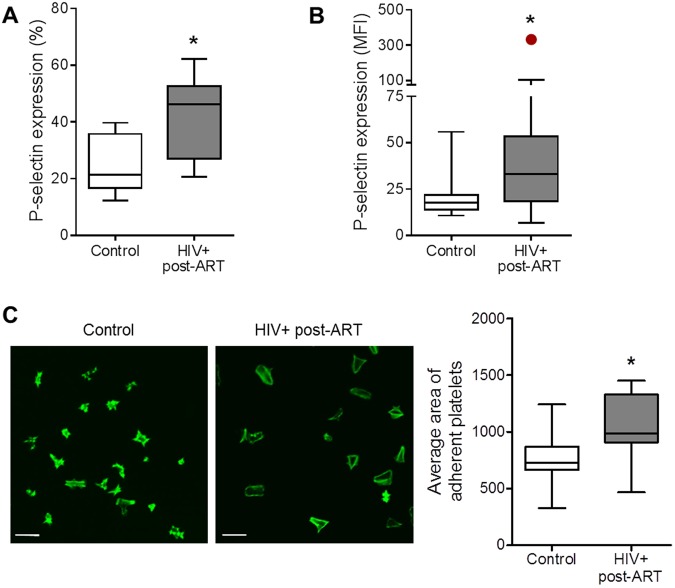
Figure 1.
Increased platelet activation in HIV-infected individuals under stable cART. (A,B) The percentage of platelets with P-selectin surface expression (A) and the mean fluorescence intensity (MFI) for P-selectin labeling on platelets (B) were assessed in freshly-isolated platelets from healthy subjects (control) or from HIV-infected individuals under viral control (undetectable HIV-1 viral load in the peripheral blood). (C) Average area of spontaneous spreading on fibrinogen-coated surfaces by platelets from control or HIV-1-infected subjects labeled with phalloidin. Images are representative of eight HIV-infected individuals and healthy volunteers analyzed in parallel. Scale bars represent 10 µm. The horizontal lines on the box plots represent the median, the boxes limits represent the interquartile ranges and the whiskers indicate 5–95 percentile. The dot in panel B represents an outlier (p < 0.05 in Grubbs’ test) that was excluded from statistical analysis. The asterisk (*) signifies p < 0.05 compared to healthy volunteers.

