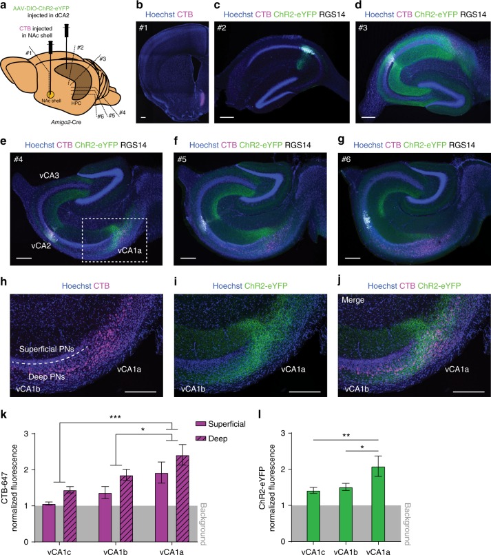
Fig. 5.
dCA2 targets a vCA1 site containing PNs that project to the NAc shell. a AAV-DIO-ChR2-eYFP was injected in dCA2 and the retrograde label CTB-647 was injected into the NAc shell of Amigo2-Cre mice. Approximate location of sections in b, c, d, e, f, and g are indicated (position #1, #2, #3, #4, #5, and #6, respectively). b Site of injection of CTB-647 (magenta). c Dorsal HPC section showing ChR2-eYFP (green) expression in dCA2 PN cell bodies (co-expressing RGS14, white) with no CTB-647 staining (lack of magenta). d Transverse section of intermediate HPC showing dCA2 fibers (labeled with ChR2-eYFP, green) distributed throughout CA3 and CA1 and sparse CTB-647 labeling (magenta). e–g Transverse sections progressing through vHPC showing dCA2 fibers (green) in vCA3, distal vCA1 (vCA1a) and adjacent ventral subiculum. Retrograde CTB-647 labeling of projections to NAc shell is seen in deep distal vCA1 (vCA1a) and adjacent ventral subiculum (magenta). h–j High magnification views of dotted area in e. k Quantification of normalized CTB-647 fluorescence in deep versus superficial PNs cell layers in vCA1c, vCA1b, and vCA1a. Ventral CA1 fluorescence was greater in deep versus superficial layers (n = 9 slices, 3 mice, repeated measures two-way ANOVA: deep/superficial F(1,8) = 55.05, P < 0.0001). Fluorescence was greater in distal (vCA1a) versus more proximal (vCA1b and vCA1c) areas (n = 9 slices, 3 mice, repeated measures two-way ANOVA: proximal-distal F(2,16) = 14.96, P = 0.0002, vCA1a vs vCA1b P = 0.0135, vCA1a vs vCA1c P = 0.0002, vCA1b vs vCA1c P = 0.1404, Tukey’s multiple comparisons test). l Quantification of normalized fluorescence intensity of ChR2-eYFP signal in dCA2 projections in vCA1. Signal was more intense in vCA1a compared to vCA1b and vCA1c (n = 9 slices, 3 mice, repeated measures one-way ANOVA: F(2,16) = 7.058, P = 0.0063, vCA1a vs vCA1b P = 0.0231, vCA1a vs vCA1c P = 0.0083, vCA1b vs vCA1c P = 0.8702, Tukey’s multiple comparisons test). Results in k and l show mean ± s.e.m. *P < 0.05; **P < 0.01; ***P < 0.001. Scale bars, 250 µm

