Fig. 8.
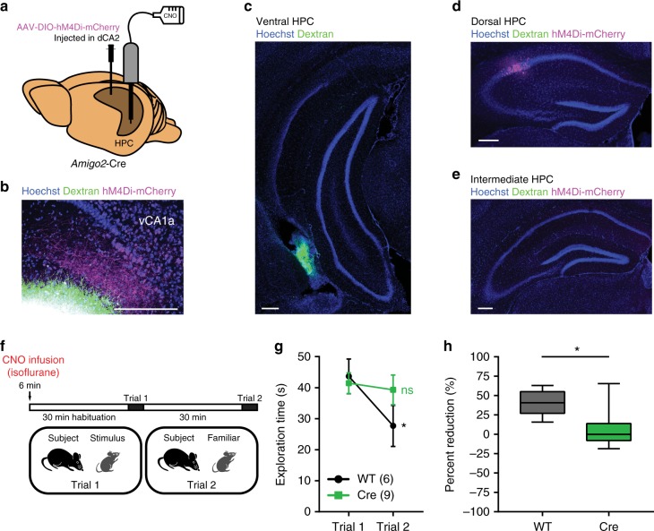
dCA2 projections to vHPC are necessary for social memory. a AAV-DIO-hM4Di-mCherry was injected in dCA2 of Amigo2-Cre mice and WT littermates. A cannula guide was implanted for local CNO or dye infusion in vCA1. b Ventral HPC horizontal slice showing overlap between dCA2 fibers located in vCA1a (labeled with hM4Di-mCherry, magenta) and dextran conjugated to Alexa Fluor 680 (green) infused in vCA1. c Ventral HPC coronal slice from an Amigo2-Cre mouse after infusion in vHPC of a dextran conjugated to Alexa Fluor 488 (green). d Dorsal HPC coronal slice from same brain as in c, showing expression of hM4Di-mCherry (magenta) but no Alexa signal. e Intermediate HPC coronal slice showing lack of signal for hM4Di-mCherry or Alexa dye. f A direct interaction test was performed 30 min after bilateral local infusion of CNO in vHPC (2 µL of 1 mM solution per side). g WT mice displayed decreased social exploration time during trial 2 (n = 6, P = 0.0110, Sidak’s multiple comparisons test) whereas hM4Di-mCherry expressing Amigo2-Cre mice showed no decrease (n = 9, P = 0.8351, Sidak’s multiple comparisons test). The two groups differed significantly (two-way ANOVA: treatment × trial F(1,13) = 4.964, P = 0.0442). h The percent reduction in interaction time in trial 2 versus trial 1 in Amigo2-Cre mice was significantly less than in WT mice (unpaired t test: t(13) = 2.962, P = 0.0110). Results in g show mean ± s.e.m. Box-whiskers plot in h present median (center line), extension from the 25th to 75th percentiles (box) and minimal and maximal values (whiskers). *P < 0.05; ns, P > 0.05. Scale bar, 250 µm (b, c, d, e)

