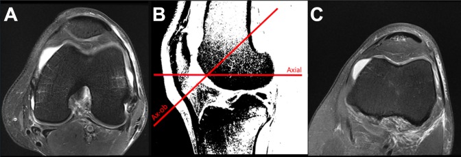
Figure 2.
Comparison of (A) standard axial versus (C) axial-oblique sequences with reference lines on the sagittal image that was used for evaluations. (B) The reference sagittal image was a single section and was converted to a poor-contrast, low-resolution image to limit potential contributions to the assessment of chondral surfaces.

