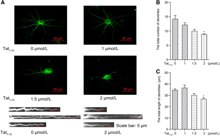
Fig. 2.
Exposure to Tat1–72 reduces the length and number of dendrites. (A) Primary cultured neurons were treated with various concentrations of Tat1–72 (0, 1, 1.5, or 2 μmol/L) for 24 h. Images were obtained by fluorescence microscopy (top). Enlarged view of individual dendritic branches (bottom). (B) Bar graph of the relative dendritic number per neuronal cell. All data are from three independent experiments. Values are presented as mean ± SEM. n = 3, *P < 0.05 versus the control group. (C) Bar graph of the relative total dendritic length per neuronal cell. H2O (2.5 μL) used as a control. All data are from three independent experiments. Values are presented as mean ± SEM. n = 3, *P < 0.05 versus the control group.

