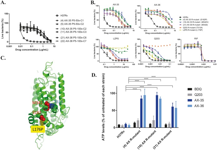
FIG 2.
Evidence of AX compounds targeting QcrB of M. tuberculosis. (A) Dose-response curves of isolated mutants resistant to either AX-35 or AX-36 compared to wild-type H37Rv. Three independent 7H9 liquid cultures of M. tuberculosis containing 50× or 100× MIC of AX-35 or AX-36 at passage 5 were plated on 7H10 medium to obtain single colonies. WGS results from AX-resistant mutants reveal mutations in QcrB. (B) Dose-response curves of AX-resistant, Q203-resistant, and LPZS-resistant mutants to AX-35, AX-36, LPZS, Q203, and RIF for cross-resistance studies. The data plotted are presented as mean ± standard deviation (SD); curves are representative of at least two independent experiments. (C) The QcrB protein of M. tuberculosis was modeled on chain A of the crystal structure of the mutant Rhodobacter sphaeroides cytochrome bc1 oxidase (PDB code 2QJK). Clustering of mutations associated with resistance to AX, Q203, and LPZS occurred around the quinol oxidation site of QcrB, indicated by the dotted blue line. (D) Intracellular ATP levels in H37Rv and AX-resistant strains were measured in the absence and presence of BDQ, Q203, AX-35, and AX-36 at 2.5× MIC after 24 h using BacTiter Glo (Promega). Data from two independent experiments are presented as mean ± SD. Statistical analysis was performed using two-way analysis of variance (ANOVA) with Tukey’s multiple-comparison test (****, P < 0.0001).

