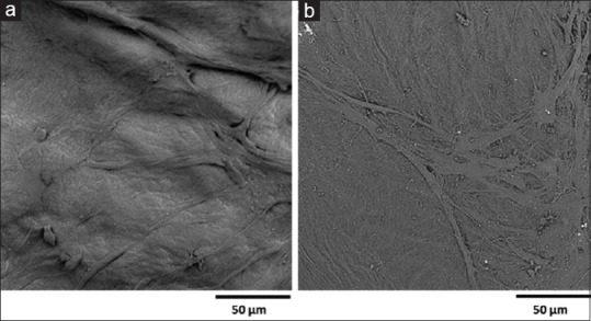
Figure 2.

Effect of different fixative material of stem cells seeded on human amniotic membrane for scanning electron microscope analysis. (a) On the specimen fixed using glutaraldehyde, the cells were not recognizable. (b) On the specimen fixed using formaldehyde, the cells were identified on the membrane surface (a and b, ×1000)
