Figure 4.

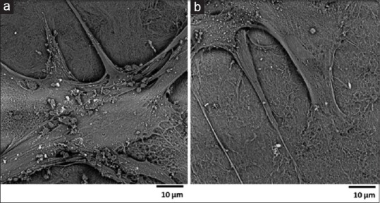
The presence of white spots on the images of specimen of stem cells seeded on human amniotic membrane of scanning electron microscope analysis (a and b, ×3000)

The presence of white spots on the images of specimen of stem cells seeded on human amniotic membrane of scanning electron microscope analysis (a and b, ×3000)