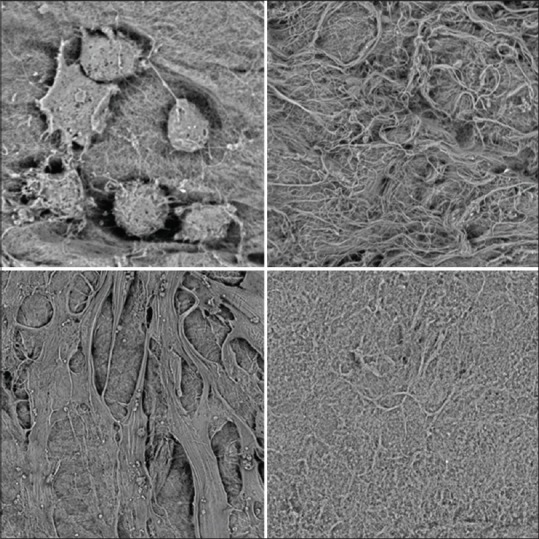
Figure 5.

Scanning electron microscope images with different magnification for human amniotic membrane samples prepared with a new protocol adjusted based on the result of the pilot study. White spots and torn layers of cells or membranes were not observed
