
Figure 2: Larvae regulate ilp2 in adults.
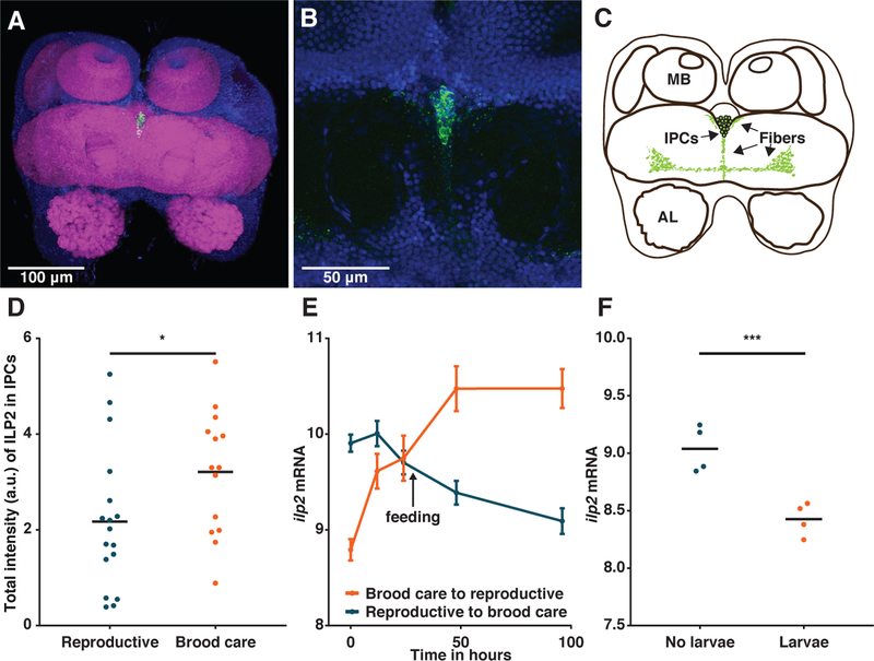
(A-C) Immunohistochemistry with anti-ILP2 antibody on an O. biroi brain localizes ILP2 peptide to a single cluster of insulin-producing cells (IPCs) in the pars intercerebralis (body-axis dorsal view). Green: anti-ILP2; blue: DAPI; magenta: phalloidin. MB: mushroom body; AL: antennal lobe. (D) Total intensity of ILP2 in the insulin-producing cells is higher in the brood care phase than in the reproductive phase (n≥14, t-test; p=0.046). (E) RNA-Seq time course shows that the addition of larvae downregulates ilp2, whereas the removal of larvae upregulates ilp2 (n≥4, time:transition interaction, Likelihood Ratio Test with 5% FDR correction; p<10−15). The black arrow indicates when ants with larvae were fed, i.e. changes in expression beyond that time point are confounded by differences in nutrition. Error bars depict SEM. Data from (28). (F) RNA-Seq on ant brains shows that under nutritionally controlled conditions, ilp2 is upregulated eight days after larvae are removed from O. biroi workers in the brood care phase (n=4, Wald test with 5% FDR correction; p<10−6). Data are variance-stabilized transformed read counts. Horizontal bars indicate means.