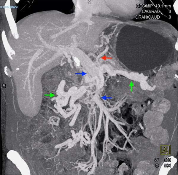
Fig. 2.
Three dimensional CT reconstruction 8 months after diagnosis, showing persistent SMV occlusion and robust venous collateralization. Blue arrows identify the portal-splenic confluence and the distal SMV, with intervening occlusion. Green arrows identify large right and left mesocolonic collaterals that communicate with the distal SMV. The red arrow identifies a dilated gastric vein that communicates between left mesocolonic collaterals and the splenic vein through retroperitoneal collaterals (not seen)

