
Figure 5:
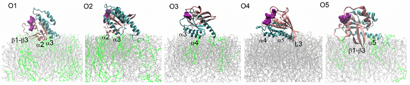
Typical orientations of K-Ras4A relative to membranes. The lobe 1 domain is colored in pink, lobe 2 and HVR in cyan, POPC in grey, POPS and PIP2 in green. The main secondary structure elements in contact with the membrane are marked. GTP nucleotide (shown as surface representation in purple) and the two switch regions are typically located at least 1.0 nm away from the membrane surface.